2024
Digital Holographic Microscope
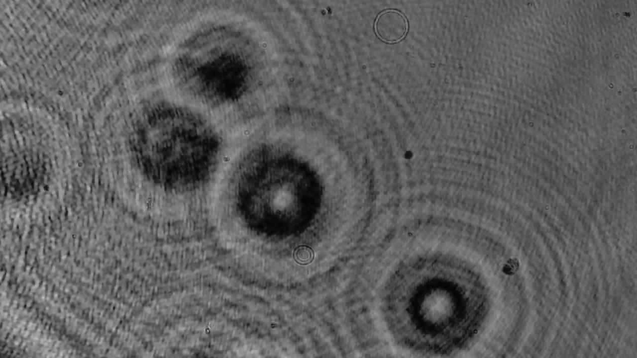
Digital Holographic Microscopy (DHM) is an advanced phase microscopy technique where a hologram is created by the interference of two light beams: the object beam and the reference beam. This method offers sharper edges, higher contrast, and multiple interference lines compared to traditional brightfield imaging.
Our work explored different DHM configurations, including:
- Machine configurations: Lens-less or lens-based setups, and inline or off-axis designs.
- Sample configurations: Testing with dry or wet, and artificial or natural samples.
We also decided to design and build our own protoype, to test the methods we developed.
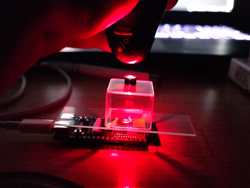
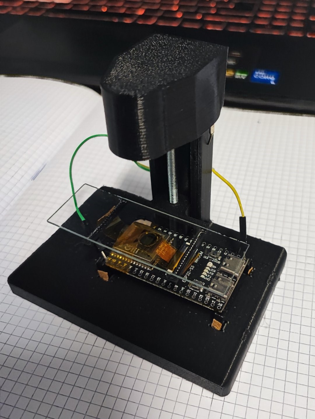
A few pictures of the first prototype:

The Team:
Project had been completed with team members Dániel Csata, Francesca Rosa Diz, Maurizio Tirabassi and Imre Vida, under the supervision of Alessandro Molani, with external contributions from Abosa Szakál and Sebestyén Nagy.
Key points of our work:
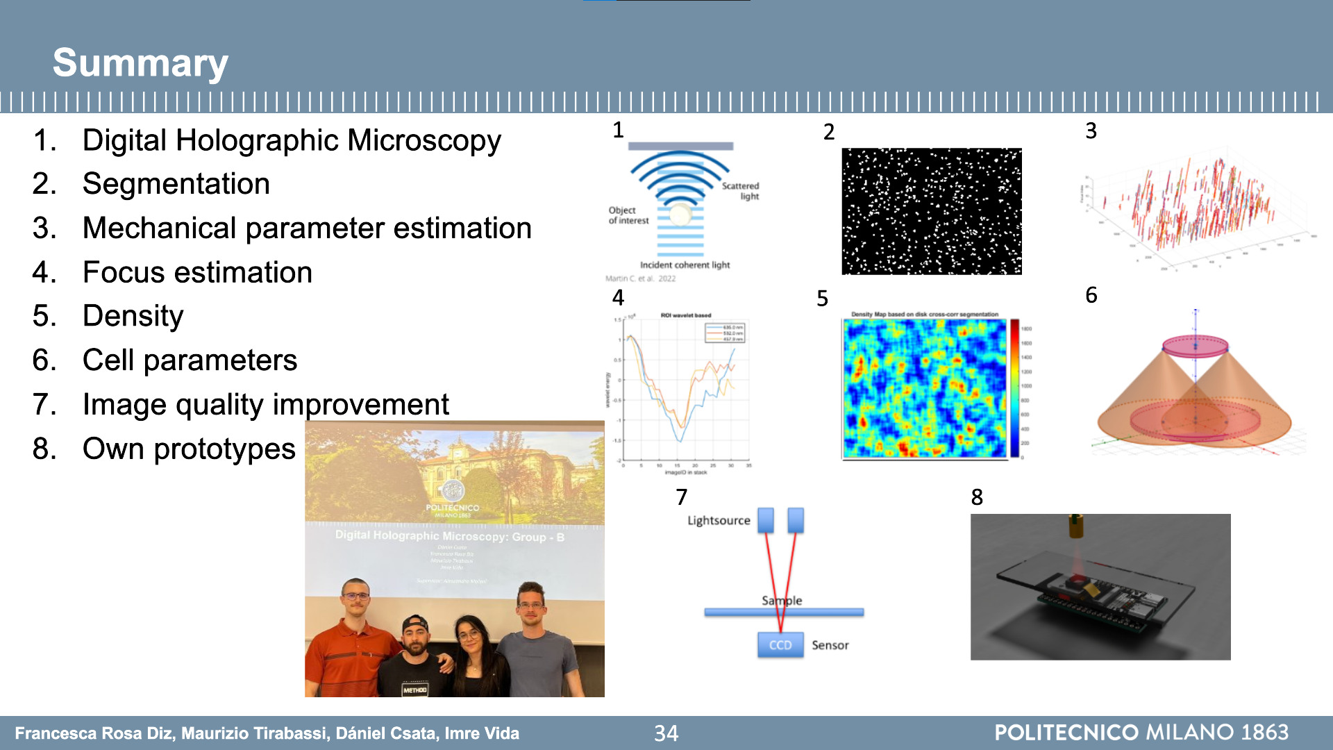
- Segmentation: Implemented edge detection, morphology techniques, and watershed transforms to isolate cells.
- Mechanical Parameter Estimation: Developed methods to correct position shifts and analyze individual cell trajectories.
- Focus Estimation: Proposed wavelet transformation-based techniques for more robust focus tracking.
- Density Mapping: Leveraged 2D and 3D convolution techniques to evaluate sample densities without the need of full image segmentation.
- AR Visualization: Developed a proof of concept augmented reality application for studying spatial cell distributions.
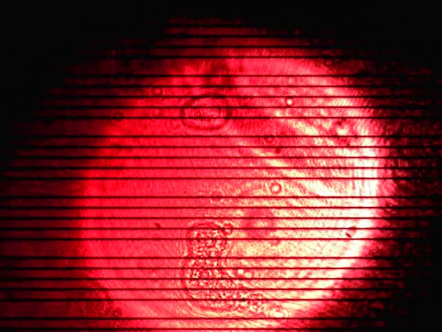
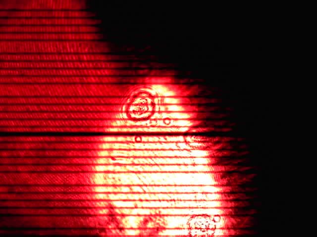
- Prototype Development:We designed and built a fully functional Digital Holographic Microscope and managed to take holographic images and videos with it.
Prototype Development:
We designed and built our own DHM prototype featuring:
- MCU: STM32S3 with USB-C and WiFi connectivity.
- Light Source: Coherent 650nm point source (modified laser diode).
- Imaging Capabilities: Variable resolution of approximately 3.07 µm/pixel with an adjustable sample holder and a modified camera.
- Future Plans: Onboard parameter estimation with embedded algorithms.
3D renderings and a printable case design were also developed for compact usability.




Summary:
Our final presentation slides: